Gastrointestinal stromal tumour (GIST’S) express somatostatin receptors Gastrointestinal stromal tumors (GISTs) start in very early forms of special cells in the wall of the GI tract called the interstitial cells of Cajal (ICCs). ICCs are sometimes called the “pacemakers” of the GI tract because they signal the muscles in the GI tract to contract to move food and liquid along. More than half of GISTs start in the stomach. Most of the others start in the small intestine, but GISTs can start anywhere along the GI tract.
51 yrs old male with h/o haematuria.
USG abdomen-Bilateral renal cyst, prostatemegaly & well Differentiated soft tissue lesion in retroperitoneum ant to IVC
MRI report showed a duodenal? Gastrointestinal stromal tumour (GIST)?? Leiomyoma. Suspicion of Neuro-endocrine tumour was high and hence ordered whole body octreotide imaging.
99mTc TEKTROTYED WHOLE BODY & SPECT CT OCTREOSCAN
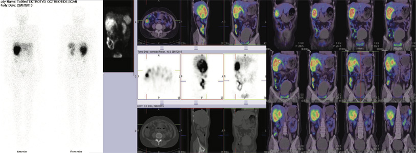
It showed isolated fairly large sized somatostatin receptor expression (SRS) lesion in III part of duodenum, which is corresponding with contrast CT/MRI images favouring GIST tumour ( as shown in pictures). Rest of the whole-body images were unremarkable.
CASE OF SMALL CELL NEUROENDOCIRNE CARCINOMA ON LIVER BIOPSY OF THE RIGHT LOBE LESION IN SEGMENT V & VI.
50 yrs old female Ref for WHOLE BODY OCTREOSCAN FOR the metastatic work up
99mTc-TEKTROTYED WHOLE BODY & SPECT CT OCTREOSCAN
It showed large somatostatin receptor expression (SRS) lesion seen in right lobe of liver involving segment V & VI. Rest of the whole-body images were unremarkable.
OPERATED CASE OF WELL DIFFERENTIATED NEURO-ENDOCRINE TUMOR (CARCINOID). REF FOR OCTREOSCAN TO LOOK FOR RESIDUAL DISEASE /METASTATIC DISEASE
55 yrs old male underwent mesenteric tumor excision (70 cms of small bowel+ Right hemicolectomy +liver lesion excision in October 2015.
99mTc-TEKTROTYED WHOLE BODY & SPECT CT OCTREOSCAN
It showed Somatostatin receptor expression (SRS) lesion seen in left adrenal gland. Rest of the whole-body images were unremarkable.
Relook CT scan showed lesion in left adrenal gland and offered surgical excision vs medical management.
WHAT IS THE CLINICAL RELEVANCE OF SOMATOSTATIN RECEPTOR SCINTIGRAPHY A positive scan predicts the suppressive effect of Somatostatin analogues on hormone secretion from endocrine active tumours.
The use of octreoscan led to a change in staging in 24% of cases and in 25% changed the planned surgical intervention.

99mTc Tektrotyed whole body and SPECT CT Octreoscan
It showed isolated fairly large sized somotostatin receptor expression (SRS) lesion in III part of duodenum, which is corresponding with contrast CT/MRI images.
Rest of the whole body images were unremarkable.

99mTc Tektrotyed whole body and SPECT CT Octreoscan
Large somatostatin receptor expression (SRS) lesion seen in right lobe of liver involving segment V & VI.
Rest of the whole body images were unremarkable.

99mTc Tektrotyed whole body and SPECT CT Octreoscan
Somatostatin receptor expression (SRS) lesion seen in left adrenal gland.
Rest of the whole body images were unremarkable.